Videoscope-Assisted Minimally Invasive Surgery (V-MIS)
Gum disease has traditionally been treated by trimming away the infected gum tissue and by re-contouring the bone.This usually results in longer, more sensitive teeth. We routinely use new and more sophisticated minimally invasive approaches that result in less discomfort and gum shrinkage compared to older surgical methods.
Until recently, this periodontal surgery was often completed using large incisions that resulted in recession and post-operative discomfort. Through the advances of a periodontal videoscope, the problems are greatly reduced.
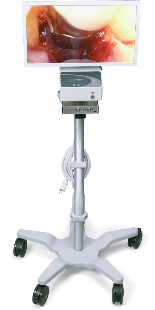
The latest and most effective device for treating periodontal disease is the dental videoscope. This device allows the use of minimally invasive procedures to remove calculus (tartar) from below the gums. A videoscope is a small camera that is inserted into the periodontal pocket and magnifies the tooth root and surrounding bone up to 50x on an external monitor to allow excellent visualization of the site. A small incision is required in order to allow the tip of a videoscope to be placed in the diseased periodontal pocket. After the root(s) are thoroughly cleaned and a biologic modifier is used to stimulate bone growth, the tissue is often closed with just a single stitch. Typically, patients have minimal to no post-operative pain, swelling, and recession due to the minimally-invasive nature of the procedure.